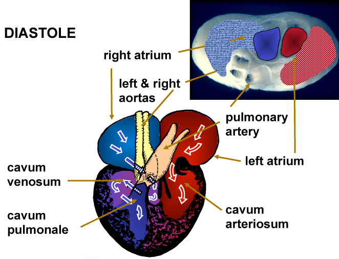
Schematic (ventral view) of blood flow in the heart of a turtle during diastole (filling phase) & photograph (cranial view) illustrating inflow and outflow tracks Oxygen-rich blood (red) returning to the heart from the lungs enters the left atrium and the left side of the ventricle, the cavum arteriosum. Oxygen-poor blood (blue) that is flowing to the heart from the rest of the body enters the right atrium and then flows into a central ventricular chamber, the cavum venosium. This blood mixes with the residual blood in the chamber, which is oxygen-rich, and then the admixture flows into the right side of the heart, the cavum pulmonale. Without this mixing, the right side of the heart would contain only the oxygen-depleted blood. After Farmer and Hicks, 2002
|
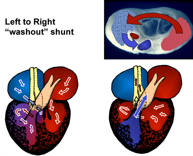
Schematic (ventral view) of blood flow in the heart of a turtle during Systole (contraction and ejection) & photograph (cranial view) showing inflow and outflow tracks The admixture in the cavum pulmonale is ejected directly into the pulmonary artery, sending the partially enriched blood back to the lung. The oxygen-rich blood in the cavum arteriosum must travel through the cavum venosum before being ejected into the left and right aortas. Thus the residual blood in this chamber at the end of systole is oxygen-rich and is the source of the left-to-right shunted blood on the next diastolic phase. After Farmer and Hicks, 2002
|
|---|---|
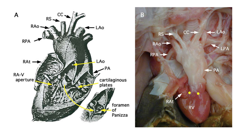
Cardiac anatomy of alligator heart and great vessels, (ventral view) A) Schematic (after Greenfield and Morrow, 1961) of the heart showing inflow into the ventricle from the right and left atria and the outflow of the left aorta and pulmonary artery. Inset shows the connection between the left and right aortas (the foramen of Panizza). (B) Photograph of this anatomy (ventral view). The ability of the animals to eject oxygen-poor blood into the LAo is created by partial closure of a cog-tooth valve in the pulmonary artery, which results in a pressure rise large enough in the right ventricle to cause blood to be ejected into the left aorta. Shunting occurs when animals are in a parasympathetic state, when in a state of excitement there is no shunting and the cardiovascular system works that that of a mammal or bird.
This outflow tract was blocked by surgical occlusion at the points marked by yellow dots (from Farmer et al. 2008). cc=common carotid, LPA=left pulmonary artery, PA=common pulmonary arterial trunk, LAo=left aorta, RAo=right aorta, RAt=right atrium, LAt= left atrium, RA-V aperture=right atrial-ventricular aperture, RPA=p.
|
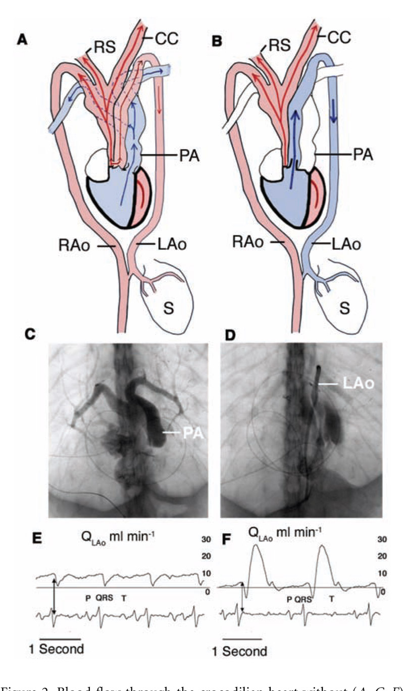
Anatomy and shunting pattern in Alligators A) Schematic of cardiac anatomy (ventral view) without an intracardiac shunt. Oxygen-rich blood (red) is ejected into the right aorta and then flows into the left aorta through the forament of Panizza. B) Schematic of flow during a shunt, oxygen-poor blood flows into the left aorta from the right ventricle. C) Angiogram with radiopaque material injected into the right ventricle along with acetylcholine to test the effectiveness of surgical occlusion. C) Angiogram with radiopaque material flowing into the left aorta from the right ventricle. E) Pattern of flow in the left aorta without a shunt. F) Flow in the left aorta during shunting. (from Farmer et al. 2008). |
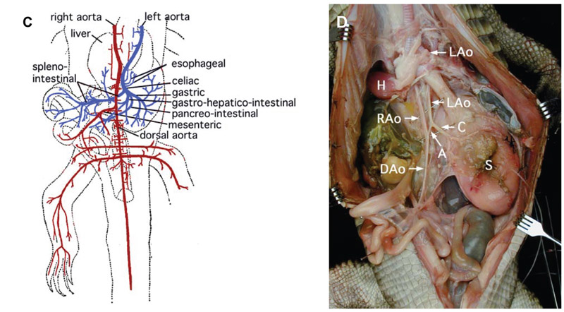 Digestive anatomy of the the American alligator, ventral view C) Schematic (after Reese 1915) and (D) photo (Farmer et al. 2008) of the left and right aortas and the arrangement of the blood vessels feeding the gastroinstinal system. When crocodilians right-to-left shunt, blood that is rich in acid and carbon dioxide (blue) flows through the left aorta to irrigate the stomach, spleen, pancreas, and small intestine. Blood to the large intestine and hindlegs and tail is an admixture from left and right aorta. A=anastomosis between left and right aortas, C=celiac, DAo=dorsal aorta, LAo=left aorta, RAo=right aorta, S=stomach. |
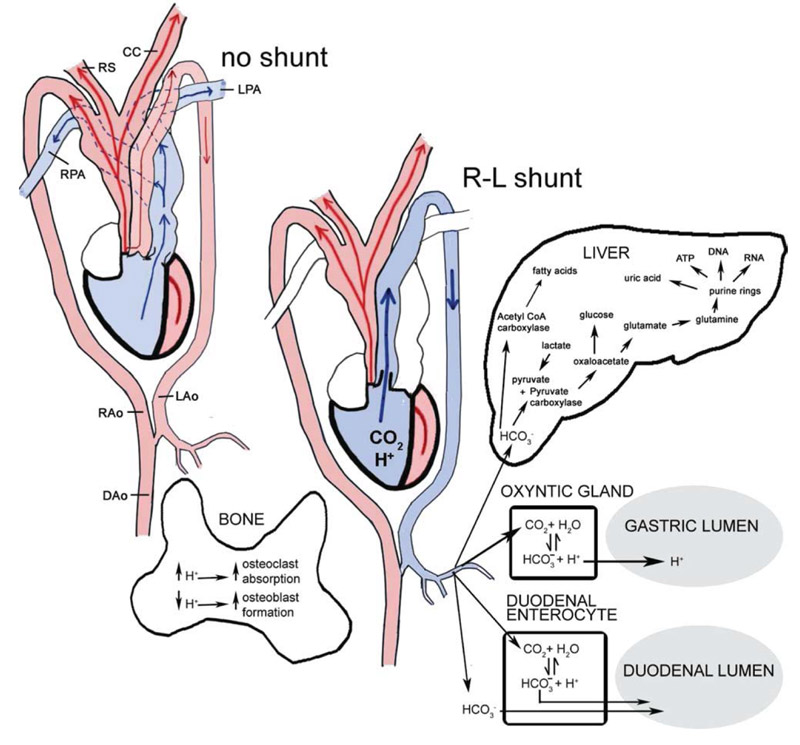 Metabolic pathways that use carbon dioxide for synthesis or that are affected by pH A number of synthetic pathways make use of carbon dioxide or bicarbonate. These include the production of gastric acid, dudoenal base, the synthesis of fatty acids, the formation of purine rings important in the production of ATP, DNA, RNA etc. Levels of acid and carbon dioxide also affect synthetic processes in the bone. After Farmer 2011 |